Estimated reading time: 4 minutes
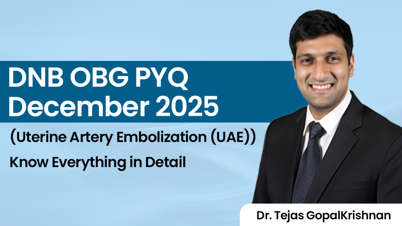
Dear residents, If you appeared for the DNB December 2025 OBG exam, this question probably caught your attention, right?
“Discuss the indications, procedure, and complications of Uterine Artery Embolization (UAE).”
Here, Dr. Tejas GopalKrishnan highlighted why this question is so important. According to him, there isn’t much left to ask beyond these three areas—indications, procedure, and complications. That makes this a topic every DNB aspirant should be comfortable with.
UAE Is Not Just About Fibroids
Most of us study uterine artery embolization while reading fibroids and often stop there. But that’s exactly where many students lose marks in exams.
UAE has both obstetric and gynecological applications.
Obstetric Indications
One of the most important indications is Placenta Accreta Spectrum (PAS). With rising cesarean section rates, PAS has become a frequently discussed topic in examinations as well as clinical practice.
Another indication is postpartum hemorrhage (PPH). Although UAE is not usually the first option in an actively bleeding unstable patient, it remains a recognized option in selected stable cases.
Cervical ectopic pregnancy is another situation where UAE can be extremely useful. Since the cervix lacks a strong contractile mechanism, surgical intervention can result in significant bleeding. Embolizing the uterine arteries beforehand helps reduce blood loss and makes management safer.
Gynecological Indications
The most commonly remembered indication is, of course, uterine fibroids.
Other important indications include:
- Adenomyosis
- Uterine arteriovenous malformations (AVMs)
- Advanced gynecological malignancies where bleeding control is required
A common mistake students make is mentioning only fibroids. In a 10-mark question, that answer remains incomplete.
How Is UAE Performed?
The procedure is usually carried out by an interventional radiologist.
The catheter is introduced through the femoral artery and guided towards the internal iliac artery. From there, the uterine arteries are identified and embolized.
An important point for exams is that both uterine arteries are embolized.
Why?
Because if only one side is embolized, collateral circulation can continue supplying the pathology, reducing the effectiveness of the procedure.
After embolization, angiography is repeated to confirm successful blockage of blood flow before removing the catheter.
Complications You Should Never Forget
A complete answer is impossible without discussing complications.
The most commonly discussed immediate complication is post-embolization syndrome, which presents with:
- Fever
- Pain
- Nausea
- Malaise
This happens because ischemic tissue releases inflammatory mediators and cytokines.
Other immediate complications include:
- Hematoma at the puncture site
- Pelvic pain and cramping
- Pseudoaneurysm formation
- Arterial dissection
- Non-target embolization
One particularly important complication is accidental embolization of the ovarian artery, which may result in ovarian dysfunction and even premature ovarian insufficiency.
What About Fertility?
This is often the final point examiners expect.
Uterine artery embolization is generally avoided in women who still plan to conceive.
Studies have linked UAE with:
- Increased miscarriage rates
- Higher risk of preterm birth
- Abnormal placentation
- Reduced fertility potential
That is why it is usually not the preferred option in younger women with fibroids who have not completed their families.
DNB Exam Takeaway
This was one of the highest-yield questions from the DNB December 2025 OBG paper. If you had covered UAE thoroughly, this question offered a great opportunity to score.
For exam writing, remember a simple formula:
Indications → Procedure → Complications → Fertility Concerns
Follow this structure, add a simple diagram, and your answer becomes much more scoring.
Want More DNB OBG PYQs Like This?
At Conceptual OBG, we don’t just discuss answers, we discuss the logic behind them and the similar types of question and what can be possible questions from that topic. From DNB PYQs and expected questions to high-yield concepts that repeatedly appear in exams, the focus is on helping you understand what really matters in the DNB Exam.
If you’re serious about DNB OBG preparation and don’t want to miss important topics, subscribe to Conceptual OBG and get access to discussions that can genuinely make a difference in your exam performance.
Watch Video: Dec DNB 2025 OBG Question Discussion with Dr. Tejas GopalKrishnan | Conceptual OBG | DNB Exam
![]()